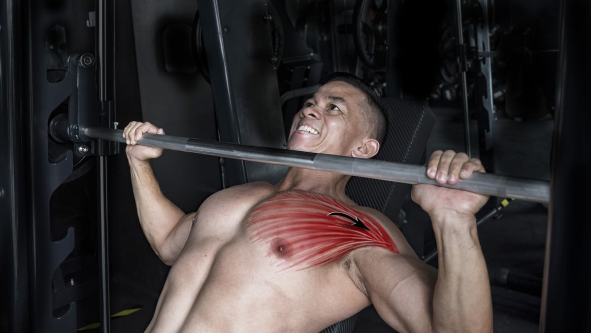
Reported injuries involving the pectoralis major muscle have significantly increased in the last 25 years. These injuries can range from contusions (bruising) and inflammation (swelling) to complete tears resulting in significant loss of function. They often cause pain, weakness, deformity to the chest wall, and can lead to noticeable changes in overall shoulder function. Military personnel, laborers, even older adults injure their pectoralis major muscle when excessive weight is lifted or moved beyond what the muscle can handle (Fig. 1). After pectoralis major injuries occur, they can be disabling, especially to athletes such as weightlifters. Several factors including recognition of the injury and timely care can help prevent delay in necessary treatment and improve functional outcomes. Treatment is dependent upon the severity of the injury, your activity level and demand, and your ability to perform activities of daily living.
Anatomy
The 2 pectoralis muscles, pectoralis major and pectoralis minor (the larger and smaller muscles of the chest), connect the front of the chest wall with the humerus (upper arm bone) and shoulder joint (Fig. 2). The pectoralis major is a thick, fan-shaped muscle consisting of 2 heads, the clavicular and sternal. The clavicular head originates from front of the clavicle (collarbone) while the sternal head arises from the sternum (breastbone) and the first through sixth ribs. The 2 portions of the muscle then converge on the outer side of the chest with the subclavius muscle (the small, triangular muscle between the clavicle and first rib) to form the axilla or armpit. The multiple origins and insertions of the pectoralis major muscle allow it to initiate a wide range of actions of the arm, enabling it to adduct (draw toward the body), flex (bend), and internally rotate (turn toward the body).
Causes of injury
Repetitive or prolonged activity can cause the tendons of the pectoralis major muscle to degenerate, resulting in a weakened tendon that is more susceptible to injury. Chronic muscle imbalances, weaknesses, tightness, and abnormal biomechanics, especially when combined with excessive training without appropriate rest, can contribute to the development of significant injury. By contrast, acute strains or tears to the pectoralis muscle occur when you place excessive tension across the muscle or tendon while it is maximally stretched or eccentrically (not central) loaded. This can occur during weight training, especially while performing a bench press (Fig. 1), chest press, or pectoral fly, and is more likely to happen when using free weights than a machine. For example, if you apply too great of an external force while the muscle is at its maximal stretch point, as during the downward movement of a bench press, it will rupture at the junction of the tendon and muscle belly. When this occurs, patients typically report hearing or feeling a pop with immediate pain.
Tears to the pectoralis major muscle can affect only a portion of the tendon or may result in a complete rupture (Fig. 3). Tears are classified as 1 of 3 grades, based on the number of muscle fibers torn and how much function has been lost, with grade 3 representing the most extensive damage. The majority of tears are grade 2.
Diagnosing the injury
During a physical examination, your doctor will look for swelling and bruising on the front of the chest that may extend to the armpit and upper arm. There may be noticeable asymmetry along the chest, and palpation (touch) can elicit pain near the armpit and the provider may even be able to palpate the tear over the chest. In addition, many patients report pain and loss of strength while pushing with the extremity. You can experience pain to the chest and front of the shoulder or armpit, but it can radiate into the upper arm or neck and may increase from an ache to a sharper pain with activity.
In the acute phase of injury, a physical exam may be difficult to perform because swelling from the injury can distort the shoulder, and pain can limit testing your strength and motion. Once the swelling has resolved, your physician can test the muscle strength while having you attempt to move your arm toward your body while he or she attempts to hold it in place. Your physician can compare the results with the opposite arm to determine the degree of limitation.
Your doctor can also use imaging to differentiate a pectoralis injury from other types of injury and to determine its extent. X-rays are often taken to look for a possible bone fragment on the tendon or other associated fractures or dislocation. Your physician may order a CT scan (computed tomography) to evaluate fractures identified on x-rays for surgical fixation or an ultrasound test that can help to assess the presence of a tear or retraction of the tendon. A MRI (magnetic resonance imaging that shows the bones, muscles, ligaments, and tendons) is often the test of choice to determine the site and extent of injury as well as giving a more advanced look at the injury to allow for possible surgical planning.
Treating the injury
The treatment for a pectoralis major injury is dependent upon the severity of injury, the extent of muscle function, and your health and general activity level. Physicians typically consider nonsurgical treatment for patients who are low demand, elderly, or have either partial tears or tears within the muscle belly. Initial management with immobilization, rest, and cold therapy followed by strengthening and stretching can offer a satisfactory to excellent functional result. Once the shoulder motion returns without pain, patients can resume daily activities. In those patients who either need to return to full strength and function or are concerned with cosmetic appearance, the physician will recommend surgical repair. Often, patients are highly satisfied with surgical repair of the pectoralis major, reporting a return of strength, structure, and overall function. Additionally, operative management can result in better function, including peak torque and overall work performance. The need for rehabilitation after surgery varies depending on how the surgeon repaired your muscle. In general, patients begin range of motion activities at 2 weeks, progressive-resistance exercises at 6 to 8 weeks with full-resistance exercises beginning at 3 to 4 months (Fig. 4). You can expect to return to your pre-injury level of function at roughly 6 months.
Returning to activity
The management of pectoralis major injuries is patient specific. In sedentary or low-demand individuals with partial or complete tears, nonsurgical management can provide acceptable to excellent results. In those who demand function and form, surgical treatment can be the best option. Although complications such as repair failure, infection, and stiffness can occur, they are relatively rare. Generally, you can return to activity and improved appearance following surgical repair with appropriate postoperative rehabilitation.
Author: Benjamin J. Catoe, DO | Columbus, Georgia
Last edited on April 10, 2024